Navigating Complex Root Anatomy with 20x Magnification
Saving a tooth with four calcified canals is only possible with microscopic visualization. Learn how we saved this tooth from the 'un-restorable' list.
Clinical Documentation: Navigating Complex Root Anatomy with 20x Magnification
When "Standard" Root Canals Aren't Enough
Every tooth is unique, and some possess anatomy that is incredibly complex. Canals can be curved like a 'C', hidden behind calcium deposits (calcified), or split into multiple tiny branches. Without advanced magnification, these hidden paths remain untreated, leading to persistent pain and eventual tooth loss.
In this case, a patient was referred to 32 Intact after a traditional root canal attempt elsewhere failed to locate two of the four canals in an upper molar.
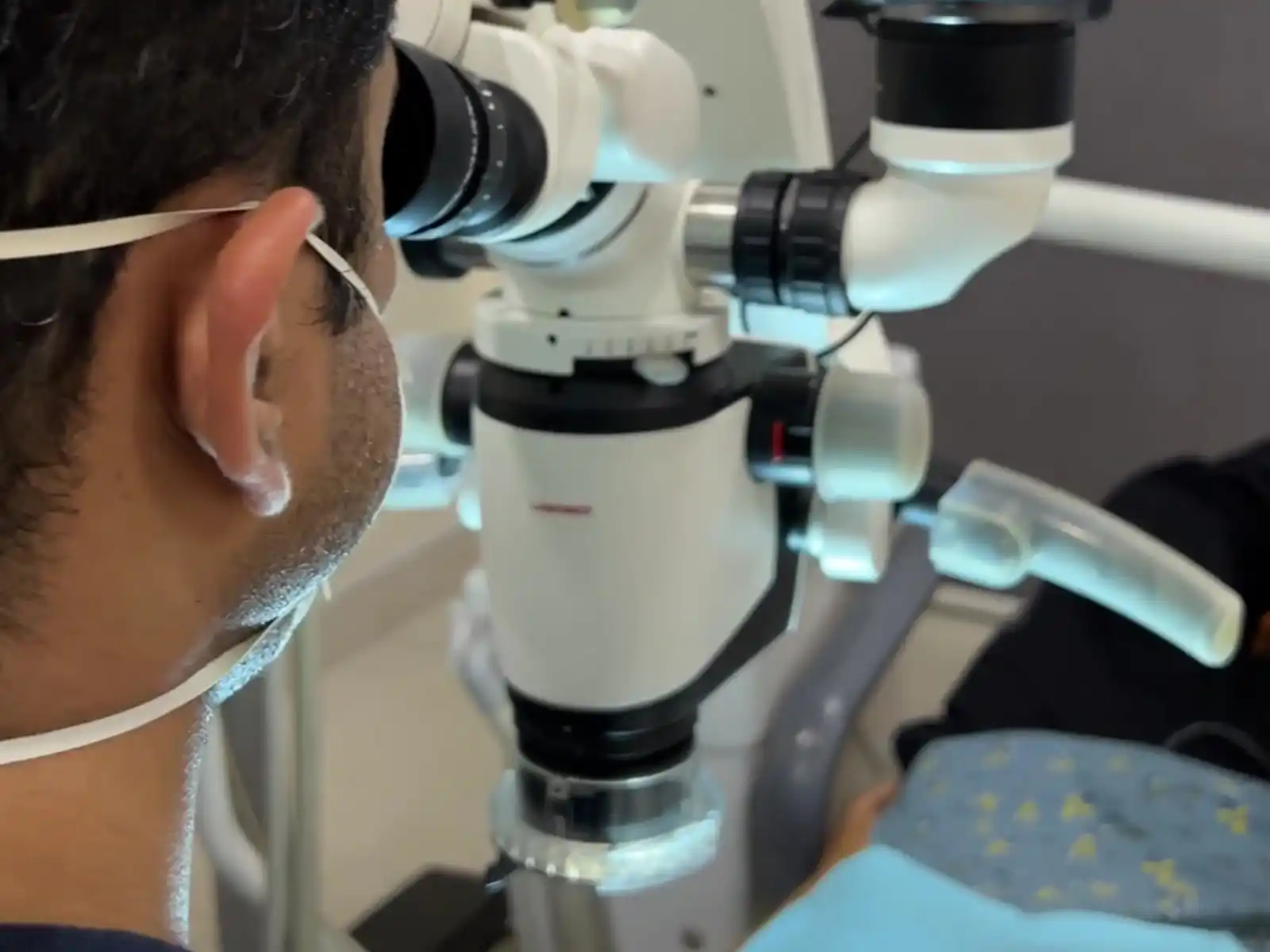
The Precision of the Dental Microscope
Under 20x magnification, what looks like a solid surface to the naked eye reveals itself to be a complex map of pathways.
1. Locating Calcified Canals: As we age or experience trauma, canals can shrink or fill with calcium. Using the dental microscope, our specialists can identify the "color change" in the dentin that signals a hidden canal.
2. Minimally Invasive Access: Because we can see exactly where we are going, we don't have to remove excess healthy tooth structure to "find" the canals. This keeps the tooth stronger for the long term.
3. Advanced Instrumentation: We use specialized, flexible Nickel-Titanium (NiTi) files that can navigate the sharp curves of the roots without breaking.
The Patient's Perspective
The difference between losing a tooth and saving it often comes down to the technology used. Our patient shared their relief:
"I was told my molar was 'impossible' to treat because the canals were too narrow to find. I was already preparing for an extraction and an expensive implant. Coming to 32 Intact was my last hope. Watching the specialist work through the microscope was fascinating—they found the 'missing' canals in minutes. It’s been a year now, and the tooth feels perfectly natural. I’m so glad I didn’t settle for pulling it out." — Verified Patient
Results: Architecture Restored
By successfully navigating all four canals, we were able to fully disinfect the tooth. The canals were then sealed with specialized fillers that adapt to the irregular shapes of the root system.
This case highlights why microscopic dentistry is the gold standard. It allows us to perform "micro-surgery" inside the tooth, preserving natural dentition and saving our patients from the more invasive and costly process of tooth replacement.